Urogenital Sphincter
Anatomy Illustrations
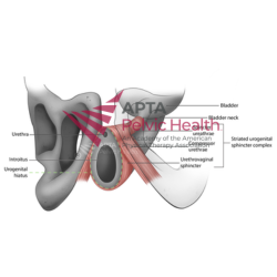
This professionally illustrated anatomical image depicts the urogenital sphincter and its associated structures within the pelvic floor. The illustration highlights the anatomical relationships involved in urinary continence, pelvic support, and coordinated voiding function.
Designed specifically for rehabilitation professionals, this illustration can be used to support patient education, clinical presentations, teaching materials, professional publications, and other non-commercial healthcare applications.
🩺 Categories
Medical Pelvic Health Urology
Ideal For
- Physical Therapists
- Pelvic Health Specialists
- Occupational Therapists
- Physicians
- Clinical Educators
- Residency Programs
- DPT and PTA course instructors
- Students
🏷️ Tags: Urogenital Sphincter · Urethral Support · Pelvic Floor Muscles · Pelvic Anatomy · Urology · Patient Education · Clinical Education · Pelvic Health
⚠️ License & Usage Terms
By purchasing this image, you agree to comply with the Academy of Pelvic Health Physical Therapy's licensing and usage policy, including all permitted uses, restrictions, attribution requirements, and other applicable terms and conditions. Unauthorized use, reproduction, distribution, modification, or failure to comply with these terms may result in immediate suspension or revocation of your license. No refunds will be issued for violations of the licensing agreement. This purchase includes a digital download only. This is not a physical product. All sales are final and non-refundable.