
Post Radical Prostatectomy
Anatomy Illustrations
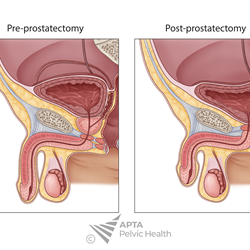
This professionally illustrated anatomical image depicts the male pelvis following radical prostatectomy, providing a clear visualization of postoperative anatomical changes. The illustration highlights removal of the prostate gland and seminal vesicles, with emphasis on the bladder neck, vesicourethral anastomosis, urethra, pelvic floor musculature, and surrounding neurovascular structures.
Designed specifically for rehabilitation professionals, this illustration can be used to support patient education, clinical presentations, teaching materials, professional publications, and other non-commercial healthcare applications.
🩺 Categories
Medical Male Pelvic Health Oncology
Ideal For
- Physical Therapists
- Pelvic Health Specialists
- Occupational Therapists
- Physicians
- Clinical Educators
- Residency Programs
- DPT and PTA course instructors
- Students
🏷️ Tags: Post Radical Prostatectomy · Prostatectomy Anatomy · Bladder Neck · Urinary Continence · Pelvic Floor Function · Prostate Removal · Surgical Anatomy · Male Pelvic Anatomy · Postoperative Recovery · Urinary Rehabilitation · Cancer Surgery · Clinical Education · Men’s Health
⚠️ License & Usage Terms
By purchasing this image, you agree to comply with the Academy of Pelvic Health Physical Therapy's licensing and usage policy, including all permitted uses, restrictions, attribution requirements, and other applicable terms and conditions. Unauthorized use, reproduction, distribution, modification, or failure to comply with these terms may result in immediate suspension or revocation of your license. No refunds will be issued for violations of the licensing agreement. This purchase includes a digital download only. This is not a physical product. All sales are final and non-refundable.